
Plik:Mixed Tumor of the Salivary Gland.jpg
Mixed_Tumor_of_the_Salivary_Gland.jpg (550 × 361 pikseli, rozmiar pliku: 33 KB, typ MIME: image/jpeg)
| Plik Mixed Tumor of the Salivary Gland.jpg znajduje się w Wikimedia Commons – repozytorium wolnych zasobów. Dane z jego strony opisu znajdują się poniżej. |
Opis
| Opis |
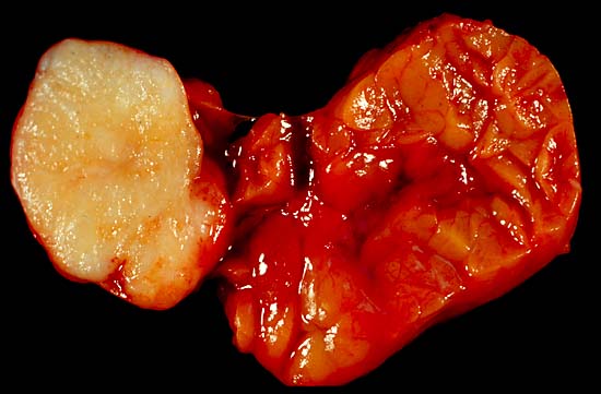
Mixed Tumor of the Salivary Gland This benign tumor of the submandibular gland, also known as pleomorphic adenoma, presented as a painless neck mass in a 40-year-old man. At the left of the image is the white tumor with its characteristic cartilaginous cut surface. To the right is the normally lobated submandibular salivary gland. Unlike most of my gross photos, this one was shot in the fresh state. This does show best what tissue looks like to the operating surgeon, but the soft, collapse-prone tissue, with its blood staining and distracting highlights, doesn't show anatomic details as well as do photos of formalin-fixed specimens. This pic was shot with a Minolta X-370 with a 100-mm Rokkor bellows lens, on Kodak Elite daylight ISO 100 film, using a blue compensator to correct for tungsten illumination. Photograph by Ed Uthman, MD. Public domain. Posted 13 May 00 |
| Źródło | http://web2.airmail.net/uthman/specimens/index.html |
| Autor | |
| Licencja (Ponowne użycie tego pliku) |
PD |
Licencja
| Ten utwór został udostępniony jako własność publiczna przez jego autora, Ed Uthman. Dotyczy to całego świata. W niektórych krajach może nie być to prawnie możliwe, jeśli tak, to: Ed Uthman zapewnia każdemu prawo do użycia tej pracy w dowolnym celu, bez żadnych ograniczeń, chyba że te ograniczenia są wymagane przez prawo.
|
Historia pliku
Kliknij na datę/czas, aby zobaczyć, jak plik wyglądał w tym czasie.
| Data i czas | Miniatura | Wymiary | Użytkownik | Opis | |
|---|---|---|---|---|---|
| aktualny | 11:51, 5 cze 2006 | | 550 × 361 (33 KB) | Patho | {{Information| |Description=Mixed Tumor of the Salivary Gland This benign tumor of the submandibular gland, also known as pleomorphic adenoma, presented as a painless neck mass in a 40-year-old man. At the left of the image is the white tumor with its ch |
Lokalne wykorzystanie pliku
Następujące strony korzystają z tego pliku:
Globalne wykorzystanie pliku
Ten plik jest wykorzystywany także w innych projektach wiki:
- Wykorzystanie na ca.wikipedia.org
- Wykorzystanie na de.wikipedia.org
- Wykorzystanie na de.wikibooks.org
- Wykorzystanie na en.wikipedia.org
- Wykorzystanie na pt.wikipedia.org
- Wykorzystanie na sr.wikipedia.org