
Plik:Schistosomiasis Life Cycle.png
Rozmiar podglądu – 752 × 600 pikseli. Inne rozdzielczości: 301 × 240 pikseli | 602 × 480 pikseli | 963 × 768 pikseli | 1280 × 1021 pikseli | 2560 × 2042 pikseli | 2936 × 2342 pikseli.
Rozmiar pierwotny (2936 × 2342 pikseli, rozmiar pliku: 1,94 MB, typ MIME: image/png)
| Plik Schistosomiasis Life Cycle.png znajduje się w Wikimedia Commons – repozytorium wolnych zasobów. Dane z jego strony opisu znajdują się poniżej. |
| Opis |
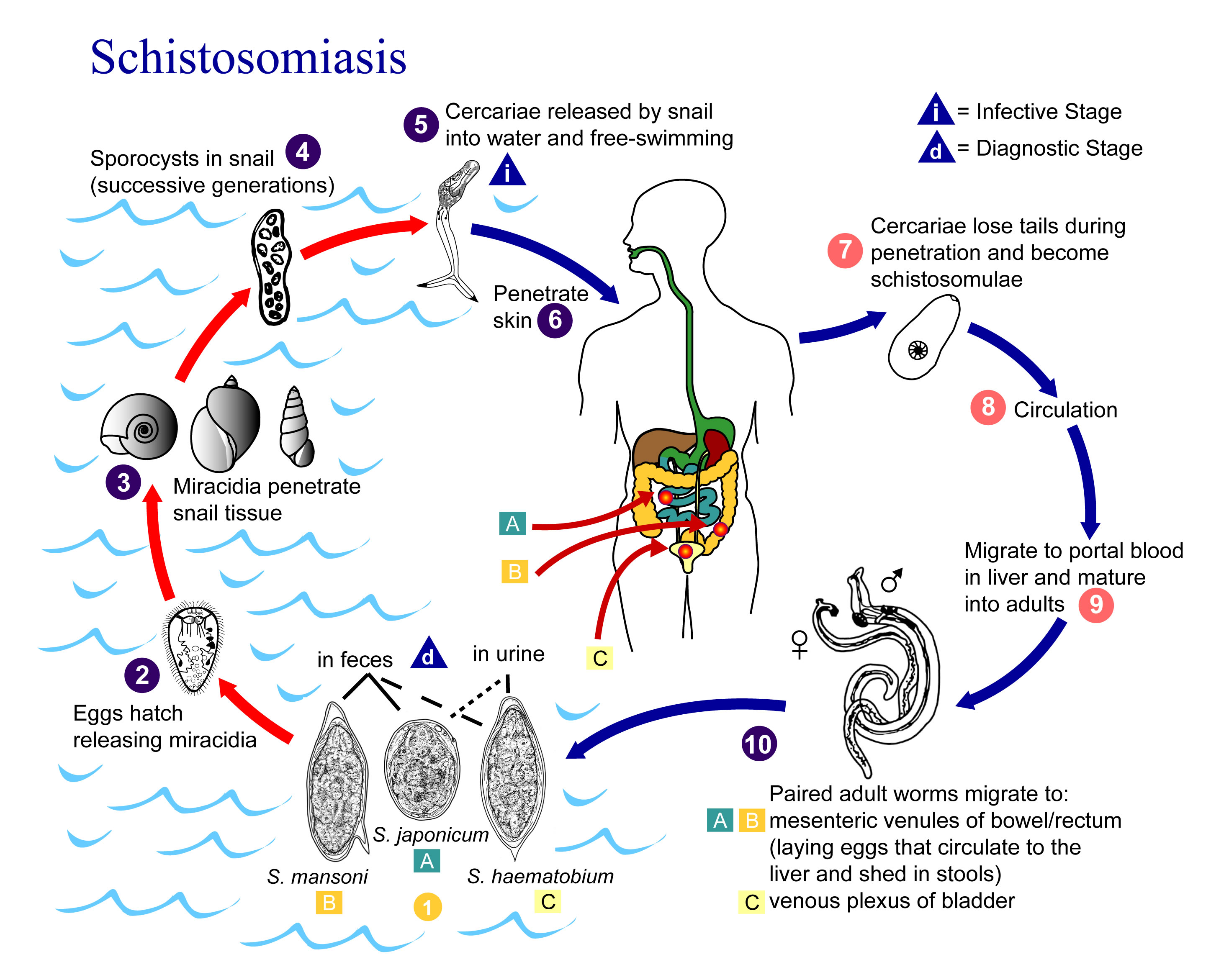
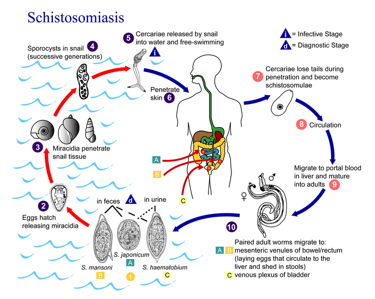
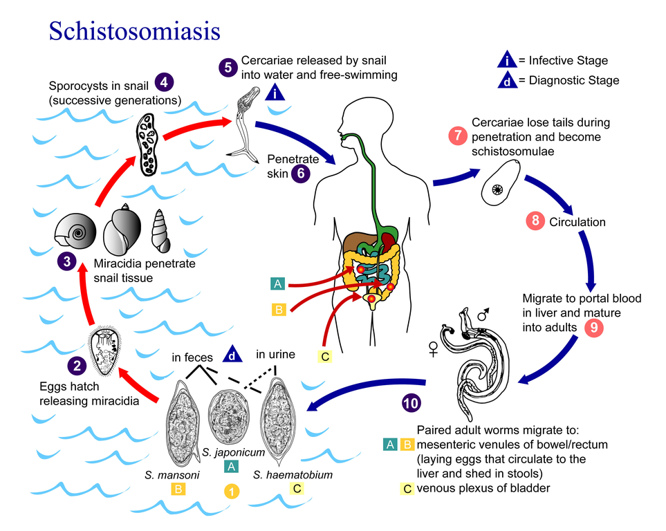
English: Eggs are eliminated with feces or urine (1). Under optimal conditions the eggs hatch and release miracidia (2), which swim and penetrate specific snail intermediate hosts (3). The stages in the snail include 2 generations of sporocysts (4) and the production of cercariae (5). Upon release from the snail, the infective cercariae swim, penetrate the skin of the human host (6), and shed their forked tail, becoming schistosomulae (7). The schistosomulae migrate through several tissues and stages to their residence in the veins (8,9). Adult worms in humans reside in the mesenteric venules in various locations, which at times seem to be specific for each species (10). For instance, S. japonicum is more frequently found in the superior mesenteric veins draining the small intestine [A], and S. mansoni occurs more often in the superior mesenteric veins draining the large intestine [B]. However, both species can occupy either location, and they are capable of moving between sites, so it is not possible to state unequivocally that one species only occurs in one location. S. haematobium most often occurs in the venous plexus of bladder [C], but it can also be found in the rectal venules. The females (size 7 to 20 mm; males slightly smaller) deposit eggs in the small venules of the portal and perivesical systems. The eggs are moved progressively toward the lumen of the intestine (S. mansoni and S. japonicum) and of the bladder and ureters (S. haematobium), and are eliminated with feces or urine, respectively (1). |
| Data | |
| Źródło | CDC DPDx |
| Autor | Nieznany |
| Inne wersje |
 |
| This image is a work of the United States Department of Health and Human Services, taken or made as part of that person's official duties. As a work of the U.S. federal government, the image is in the public domain. | 
|
Oryginalny rejestr przesyłania
This image is a derivative work of the following images:
- File:Schistosomiasis_Life_Cycle.jpeg licensed with PD-USGov-HHS
- 2009-09-02T05:15:25Z Gzuckier 3150x2400 (697710 Bytes) same pic, higher resolution
- 2005-07-10T00:44:22Z Salvadorjo 700x533 (62433 Bytes) Life cycle of schistosomiasis parasite. US Federal Government public domain archive image. Source: CDC {{PD}} [[Category:Schistosoma]]
Uploaded with derivativeFX
Historia pliku
Kliknij na datę/czas, aby zobaczyć, jak plik wyglądał w tym czasie.
| Data i czas | Miniatura | Wymiary | Użytkownik | Opis | |
|---|---|---|---|---|---|
| aktualny | 23:05, 21 wrz 2010 | | 2936 × 2342 (1,94 MB) | Leyo | {{Information |Description={{en|Eggs are eliminated with feces or urine (1). Under optimal conditions the eggs hatch and release miracidia (2), which swim and penetrate specific snail intermediate hosts (3). The stages in the snail include 2 generations o |
Lokalne wykorzystanie pliku
Poniższa strona korzysta z tego pliku:
Globalne wykorzystanie pliku
Ten plik jest wykorzystywany także w innych projektach wiki:
- Wykorzystanie na ar.wikipedia.org
- Wykorzystanie na arz.wikipedia.org
- Wykorzystanie na ceb.wikipedia.org
- Wykorzystanie na cs.wikipedia.org
- Wykorzystanie na de.wikibooks.org
- Wykorzystanie na en.wikipedia.org
- Wykorzystanie na es.wikipedia.org
- Wykorzystanie na fr.wikipedia.org
- Wykorzystanie na hu.wikipedia.org
- Wykorzystanie na hu.wikibooks.org
- Wykorzystanie na ml.wikipedia.org
- Wykorzystanie na nl.wikipedia.org
- Wykorzystanie na sv.wikipedia.org
- Wykorzystanie na uz.wikipedia.org
- Wykorzystanie na vi.wikipedia.org