
Plik:1gwe antipar betaSheet both.png

Rozmiar podglądu – 800 × 440 pikseli. Inne rozdzielczości: 320 × 176 pikseli | 640 × 352 pikseli | 1024 × 563 pikseli | 1280 × 704 pikseli | 2000 × 1100 pikseli.
Rozmiar pierwotny (2000 × 1100 pikseli, rozmiar pliku: 1,04 MB, typ MIME: image/png)
| Plik 1gwe antipar betaSheet both.png znajduje się w Wikimedia Commons – repozytorium wolnych zasobów. Dane z jego strony opisu znajdują się poniżej. |
Opis
| Opis |
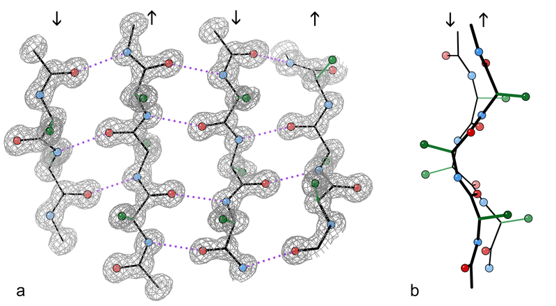
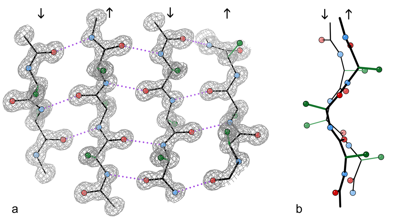
English: An example of a 4-stranded antiparallel β sheet fragment from a crystal structure of the enzyme catalase (PDB file 1GWE at 0.88 Å resolution). a) Front view, showing the antiparallel hydrogen bonds (dotted) between peptide NH and CO groups on adjacent strands. Arrows indicate chain direction, and electron density contours outline the non-H atoms. O atoms are red balls, N atoms are blue, and H atoms are omitted for simplicity; sidechains are shown only out to the first sidechain C atom (green). b) Edge-on view of the central two β strands in a, showing the righthanded twist and the pleat of Cαs and sidechains that alternately stick out in opposite directions from the sheet.
Français : Exemple d'un fragment de feuillet β à quatre chaines antiparallèles extrait de la structure cristalline de l'enzyme catalase (résolution 0,88 Å). a) Vue de face, montrant les liaisons hydrogènes (en pointillés) entre les groupes NH et CO des acides aminés adjacents. Les flèches indiquent l'orientation des chaines, et les contours de densité d'électron entourent les atomes autres que l'hydrogène. Les atomes d'oxygène sont donnés en rouge, ceux d'azote en bleu. Les atomes d'hydrogène sont omis pour plus de simplicité. Dans le même but, seul le premier carbone des radicaux est montré (en vert). b)vue par côté des deux chaines centrales montrant la torsion à droite des chaines l'une par rapport à l'autre, ainsi que les plis de chacune d'elle qui orientent les carbones portant les radicaux des acides aminés alternativement de part et d'autre de celles-ci. |
| Data | |
| Źródło | Praca własna |
| Autor | Dcrjsr |
Licencja
Ja, właściciel praw autorskich do tego dzieła, udostępniam je na poniższej licencji
Ten plik udostępniony jest na licencji Creative Commons Uznanie autorstwa 3.0.
- Wolno:
- dzielić się – kopiować, rozpowszechniać, odtwarzać i wykonywać utwór
- modyfikować – tworzyć utwory zależne
- Na następujących warunkach:
- uznanie autorstwa – musisz określić autorstwo utworu, podać link do licencji, a także wskazać czy utwór został zmieniony. Możesz to zrobić w każdy rozsądny sposób, o ile nie będzie to sugerować, że licencjodawca popiera Ciebie lub Twoje użycie utworu.

|
Ta grafika została oceniona wg kryteriów dla wartościowych grafik i jest uważana za najbardziej wartościową grafikę na Commons w zakresie: (ang.) Protein sheets and strands. Nominacja znajduje się na stronie Commons:Valued image candidates/1gwe antipar betaSheet both.png. |
Historia pliku
Kliknij na datę/czas, aby zobaczyć, jak plik wyglądał w tym czasie.
| Data i czas | Miniatura | Wymiary | Użytkownik | Opis | |
|---|---|---|---|---|---|
| aktualny | 18:00, 10 kwi 2010 | | 2000 × 1100 (1,04 MB) | Dcrjsr | {{Information |Description={{en|1=An example of a 4-stranded antiparallel β sheet fragment from a crystal structure of the enzyme catalase (PDB file 1GWE at 0.88Å resolution). a) Front view, showing the antiparallel hydrogen bonds (dotted) between pepti |
Lokalne wykorzystanie pliku
Poniższa strona korzysta z tego pliku:
Globalne wykorzystanie pliku
Ten plik jest wykorzystywany także w innych projektach wiki:
- Wykorzystanie na ar.wikipedia.org
- Wykorzystanie na bg.wikipedia.org
- Wykorzystanie na bs.wikipedia.org
- Wykorzystanie na ca.wikipedia.org
- Wykorzystanie na en.wikipedia.org
- Wykorzystanie na en.wikibooks.org
- Wykorzystanie na fa.wikipedia.org
- Wykorzystanie na fr.wikipedia.org
- Wykorzystanie na gl.wikipedia.org
- Wykorzystanie na ja.wikipedia.org
- Wykorzystanie na mk.wikipedia.org
- Wykorzystanie na ru.wikipedia.org
- Wykorzystanie na sh.wikipedia.org
- Wykorzystanie na sr.wikipedia.org
- Wykorzystanie na tr.wikipedia.org