
Plik:Human brain right dissected lateral view description.JPG
Human_brain_right_dissected_lateral_view_description.JPG (653 × 413 pikseli, rozmiar pliku: 40 KB, typ MIME: image/jpeg)
| Plik Human brain right dissected lateral view description.JPG znajduje się w Wikimedia Commons – repozytorium wolnych zasobów. Dane z jego strony opisu znajdują się poniżej. |
Opis
| Opis |
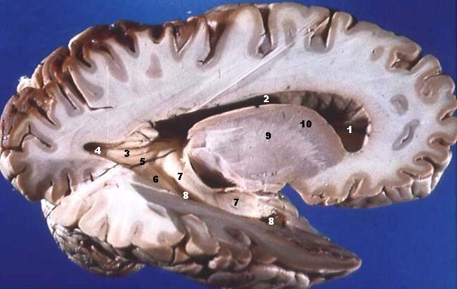
Human brain right dissected lateral view description.JPG Lateral Portion of Frontal, Parietal, Occipital, and Superior Portion of Temporal Lobe Resected. The anterior horn of the lateral ventricle is located in the frontal lobe. The body of the lateral ventricle continues posteriorly into the parietal lobe, the posterior horn into the occipital lobe, and the inferior horn down into the temporal lobe. Some structures produce elevations or bumps in the walls of the posterior and/or inferior horns of the lateral ventricles.
|
| Data | |
| Źródło | http://www.healcentral.org/healapp/showMetadata?metadataId=40566 (Internet Archive of file description page) |
| Autor |
John A Beal, PhD Dep't. of Cellular Biology & Anatomy, Louisiana State University Health Sciences Center Shreveport |
| Licencja (Ponowne użycie tego pliku) |
CC-BY |
| Inne wersje | http://commons.wikimedia.org/wiki/Image:Human_brain_right_dissected_lateral_view.JPG |
Licencja
- Wolno:
- dzielić się – kopiować, rozpowszechniać, odtwarzać i wykonywać utwór
- modyfikować – tworzyć utwory zależne
- Na następujących warunkach:
- uznanie autorstwa – musisz określić autorstwo utworu, podać link do licencji, a także wskazać czy utwór został zmieniony. Możesz to zrobić w każdy rozsądny sposób, o ile nie będzie to sugerować, że licencjodawca popiera Ciebie lub Twoje użycie utworu.
Ten plik, opublikowany pierwotnie w serwisie https://web.archive.org/web/20110514023714/http://www.healcentral.org/healapp/showMetadata?metadataId=40566, został sprawdzony 1 listopada 2013 przez administratora lub redaktora grafik Avenue, który stwierdził, że jest on dostępny w tymże serwisie pod powyższą licencją.
|
| Adnotacje | To zdjęcie jest opatrzone adnotacją: Zobacz adnotacje w Commons |
Historia pliku
Kliknij na datę/czas, aby zobaczyć, jak plik wyglądał w tym czasie.
| Data i czas | Miniatura | Wymiary | Użytkownik | Opis | |
|---|---|---|---|---|---|
| aktualny | 16:29, 22 cze 2006 | | 653 × 413 (40 KB) | Patho | {{Information| |Description='''Human brain right dissected lateral view description.JPG''' Lateral Portion of Frontal, Parietal, Occipital, and Superior Portion of Temporal Lobe Resected. The anterior horn of the lateral ventricle is located in the fro |
Lokalne wykorzystanie pliku
Następujące strony korzystają z tego pliku:
Globalne wykorzystanie pliku
Ten plik jest wykorzystywany także w innych projektach wiki:
- Wykorzystanie na ar.wikipedia.org
- Wykorzystanie na bn.wikipedia.org
- Wykorzystanie na bs.wikipedia.org
- Wykorzystanie na da.wikipedia.org
- Wykorzystanie na de.wikipedia.org
- Wykorzystanie na de.wikibooks.org
- Wykorzystanie na en.wikipedia.org
- Wykorzystanie na en.wikibooks.org
- Wykorzystanie na en.wiktionary.org
- Wykorzystanie na es.wikipedia.org
- Wykorzystanie na eu.wikipedia.org
- Wykorzystanie na fa.wikipedia.org
- Wykorzystanie na fr.wikipedia.org
- Wykorzystanie na fr.wikibooks.org
- Wykorzystanie na gl.wiktionary.org
- Wykorzystanie na he.wikipedia.org
- Wykorzystanie na he.wiktionary.org
- Wykorzystanie na hy.wikipedia.org
- Wykorzystanie na it.wikipedia.org
- Wykorzystanie na ja.wikipedia.org
Pokaż listę globalnego wykorzystania tego pliku.