
Plik:Tuberculosis-x-ray.jpg
Grafika w wyższej rozdzielczości nie jest dostępna.
Tuberculosis-x-ray.jpg (700 × 542 pikseli, rozmiar pliku: 32 KB, typ MIME: image/jpeg)
| Plik Tuberculosis-x-ray.jpg znajduje się w Wikimedia Commons – repozytorium wolnych zasobów. Dane z jego strony opisu znajdują się poniżej. |
Opis
| Opis |
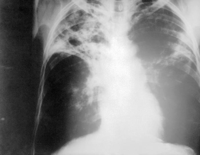
English: An anteroposterior X-ray of a patient diagnosed with advanced bilateral pulmonary tuberculosis. This AP X-ray of the chest reveals the presence of bilateral pulmonary infiltrate, and „caving formation“ present in the right apical region. The diagnosis is far-advanced tuberculosis.
Deutsch: Eine Röntgenaufnahme im anterior-posterioren Strahlengang eines Patienten, bei dem eine beidseitige Lungentuberkulose festgestellt wurde. Diese Thorax-Aufnahme zeigt beidseitige Lungeninfiltrate und eine sogenannte „Kaverne“ im rechten Oberfeld. Sie entspricht der Diagnose einer fortgeschrittenen Lungentuberkulose. |
||
| Data |
|
||
| Źródło |
|
||
| Autor |
|
||
| Licencja (Ponowne użycie tego pliku) |
PD-USGov-HHS-CDC English: None - This image is in the public domain and thus free of any copyright restrictions. As a matter of courtesy we request that the content provider be credited and notified in any public or private usage of this image. |
Licencja
Ta grafika została utworzona przez pracownika Centrum Zwalczania i Zapobiegania Chorób będącego częścią Ministerstwa Zdrowia i Usług Społecznych podczas wykonywania czynności służbowych. Jako utwór Rządu Federalnego Stanów Zjednoczonych, grafika ta znajduje się w domenie publicznej.
|
Oryginalny rejestr przesyłania
Oryginalna strona opisu była tutaj. Wszystkie poniższe nazwy użytkowników odwołują się do en.wikipedia.
- 2005-06-05 14:21 Rsabbatini 700×542×8 (32649 bytes) An anteroposterior [[X-ray]] of a patient diagnosed with advanced bilateral [[lung|pulmonary]] [[tuberculosis]]. This AP X-ray of the [[chest]] reveals the presence of bilateral pulmonary infiltrate, and “caving formation” present in the right apical
Historia pliku
Kliknij na datę/czas, aby zobaczyć, jak plik wyglądał w tym czasie.
| Data i czas | Miniatura | Wymiary | Użytkownik | Opis | |
|---|---|---|---|---|---|
| aktualny | 14:19, 20 cze 2006 | | 700 × 542 (32 KB) | Der Lange | {{Information |Description= *'''en''': An anteroposterior X-ray of a patient diagnosed with advanced bilateral pulmonary tuberculosis. This AP X-ray of the chest reveals the presence of bilateral pulmonary infiltrate, and „caving formation“ present in |
Lokalne wykorzystanie pliku
Następujące strony korzystają z tego pliku:
Globalne wykorzystanie pliku
Ten plik jest wykorzystywany także w innych projektach wiki:
- Wykorzystanie na ar.wikipedia.org
- Wykorzystanie na en.wikipedia.org
- Wykorzystanie na en.wikiversity.org
- Wykorzystanie na lt.wikipedia.org